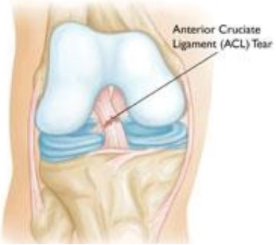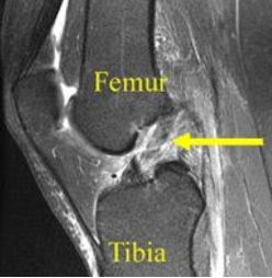
Knee Joint Showing ACL Tear
Overview of ACL Injury
Anterior Cruciate Ligament (ACL) is cord like tissue attached to bone on either side inside knee joint passing from top of shin bone (Tibia) to lower aspect of thigh bone (femur). Anterior –front Cruciate – Cros. Its function is to give stability to knee joint and prevent sliding movement between flat surfaces of tibia and femur. So thereby it also protects menisci and cartilage from shear forces
- Sports- involving sudden change in position (cutting movements)or sudden stop (deceleration) Female athletes (with very lax joint) involved in pivoting movements are prone for ACL injury.
- Trivial trauma– Twisting injury in especially overweight person with lack of balancing sense.
- Motor Vehicle accident – Motor bike accident, or a direct blow to shin bone (hyper-extension).
- There will be sudden pop sensation at the time of injury.
- Followed by swelling, painful movements.
- sometimes you may feel of giving-way sensation.
Are you an athlete who sustained ACL injury or ligament tear?
You can consult Dr. Abhijit Ranaware (Knee and sports injury Specialist)
Book Appointment by Filling Form Below:
How will be the ACL injury diagnosed?
Dr. Abhijit Ranaware will diagnose ACL tear clinically by doing specific test and then advise investigations.
- x rays – to see for over alignment of limb axis and knee joint.
- Magnetic resonance imaging (MRI) scan – will confirm the ACL tear and also reveal any associated meniscus tear, cartilage lesions which is usually seen in more than half of the ACL injury.

