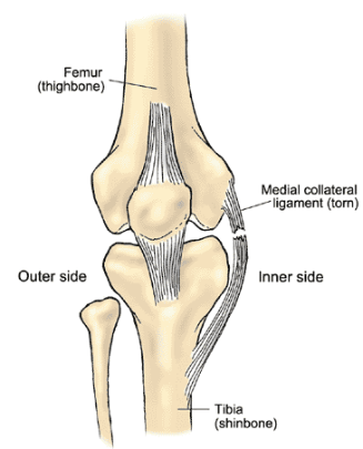
Knee joint showing medial collateral ligament (MCL) tear
Overview of MCL (Knee)
MCL injury is most common injury on inner aspect of knee. Medial Collateral Ligament that spans on medial (inner) aspect of the knee joint extends from lower thigh bone (femur) to upper shin bone (tibia). MCL is main supporting ligament and prevents inward buckling of knee. As this ligament is not inside joint most of the time needs open surgery for repair. MCL formed by three parts : Superficial MCL, Deep MCL and Posterior Oblique Ligament.
Grade of injury
I MCL sprain
II Partial tear of MCL
III Complete tear of MCL
Isolated MCL injury occurs when extreme outward force is applied inner aspect of knee or knock/ hard hit over inside of knee joint as in Motor bike accident
Contact Sports - with awkward landing on leg with twisting of knee, strong tackle with outward moving force on leg.
Severity of injury depends part of torn MCL, superficial, deep and posterior oblique. Pain, swelling and sometimes bruise on inner aspect of knee. If deep MCL is also torn patient may feel knee gives-way while weight bearing.
Knee becomes more unstable if MCL tear is associated with other ligament injury (Complex / Multiple ligament injury )
Are you a contact athlete who sustained MCL injury or knee feels unstable?
You can consult Dr. Abhijit Ranaware (Knee and sports injury Specialist)
Book Appointment by Filling Form Below:
Diagnosis
Dr. Abhijit Ranaware will examine patient in his Clinic and do specific test to diagnose MCL tear. There will be pain on palpation at the attachment of MCL and medial joint opening of knee joint with stress applied to leg. He will also evaluate to know if any associated cruciate ligament injury.
After examination he will advise :
X rays – Stress views to check amount of gapping over inside of knee joint, compared to normal side.
MRI – to confirm MCL tear and to reveal associated soft tissue injury or other ligament injury.

