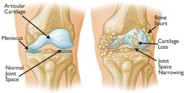
Left) Normal knee joint: Right) Knee joint with changes of Osteoarthritis
Overview of Osteoarthritis (Knee)
Osteoarthritis in Young Active individuals. Knee is largest hinge type of joint in body. It is formed by lower end of femur (thigh bone) and upper end of tibia (shin bone) and the patella (kneecap).
Articular cartilage is firm slippery tissue covering and cushioning the bony ends that form knee joint Knee joint cartilage provides smooth gliding movements of bones over each other. The lining of the knee joint capsule is called the synovial membrane which releases a fluid that lubricates cartilage and reduced friction during knee movements.
Two wedge-shaped modified cartilage tissue called meniscus are placed in thigh and shin bone those act like a “shock absorber”. They help to cushion the joint and keep knee stable.
How osteoarthritis occurs:
It is gradual process of aging or degeneration. When the cartilage wears off it becomes frayed and rough. The articular cartilage may break down over the time and the underlying bone is exposed. Exposed bones rub on each other while movement and is painful. To compensate for the lost cartilage the damaged bones may start to grow outward and form painful bony spurs.
Pain and stiffness are common symptoms.|
Pain is more on activity like walking.
Risk factors to develop osteoarthritis are :
Age - > 50 years are as risk , some time younger may affect:
- Over Weight – more the weight more stess is put on knee joints
- Heredity.
- Previous Knee injury or fractures involving articular surface.
- Systemic joint disease like Rheumatoid, Gout.
Are you suffering from knee osteoarthritis and want to know treatment options to preserve your natural knee joint?
You can consult Dr. Abhijit Ranaware (Knee and sports injury Specialist)
Book Appointment by Filling Form Below:
Diagnosis of Osteoarthritis

Knee joint X-ray Left) Normal ; Right ) with changes of Osteoarthritis (reduced joint space)

